Figure 1. [The normal human retina fundus]. - Webvision - NCBI
Por um escritor misterioso
Last updated 03 junho 2024
![Figure 1. [The normal human retina fundus]. - Webvision - NCBI](https://www.ncbi.nlm.nih.gov/books/NBK554706/bin/Archetecture_Fovea-Image006.jpg)
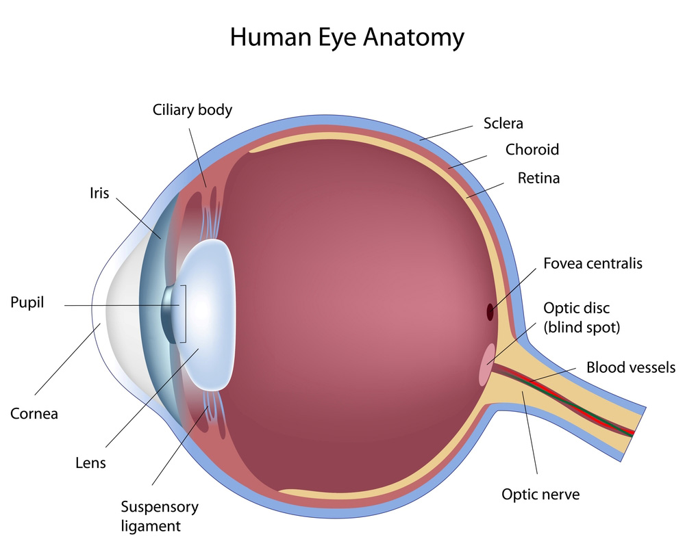
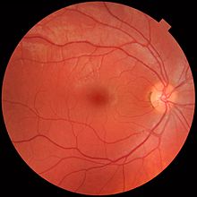
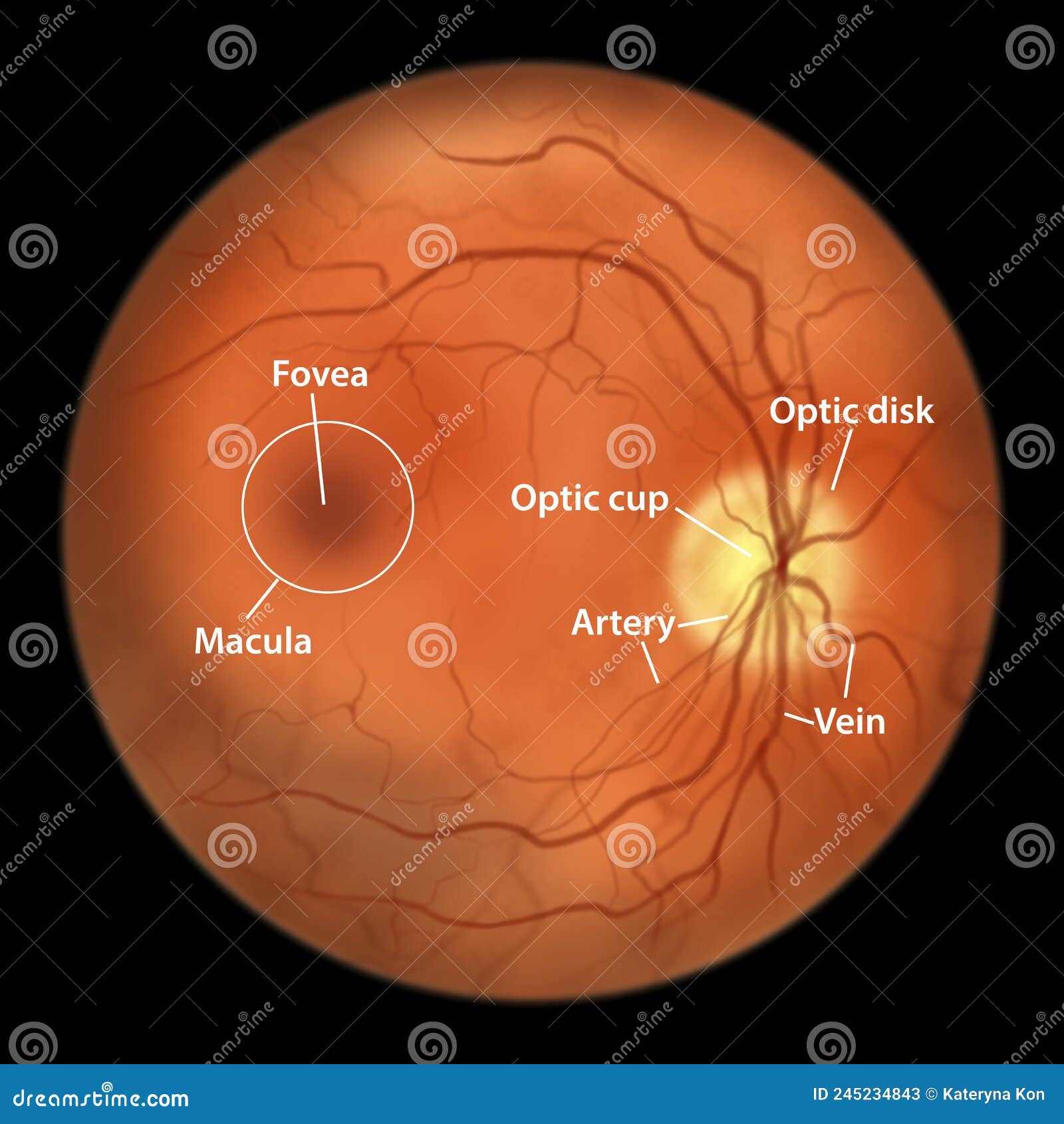
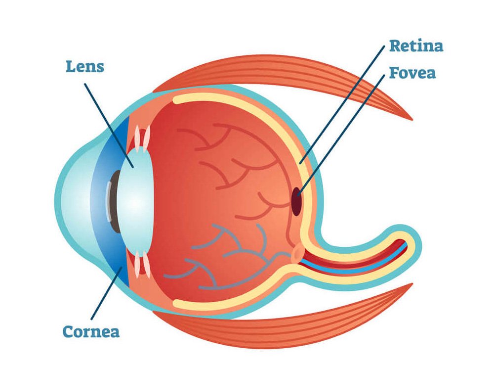
The normal human retina fundus photo shows the optic nerve (right), blood vessels and the position of the fovea (center).
![Figure 1. [The normal human retina fundus]. - Webvision - NCBI](https://media.springernature.com/full/springer-static/image/art%3A10.1038%2Fs41467-019-12917-9/MediaObjects/41467_2019_12917_Fig1_HTML.png)
Single-nuclei RNA-seq on human retinal tissue provides improved transcriptome profiling
a) Normal fundus image. b) Pathology fundus image. c) Segmentation of
![Figure 1. [The normal human retina fundus]. - Webvision - NCBI](https://webvision.med.utah.edu/wp-content/uploads/2018/07/Fig-14-macula-lutea.jpg)
Simple Anatomy of the Retina by Helga Kolb – Webvision
![Figure 1. [The normal human retina fundus]. - Webvision - NCBI](https://www.pnas.org/cms/10.1073/pnas.2307380120/asset/a3533755-1d49-4826-ba92-7697defec4a7/assets/images/large/pnas.2307380120fig08.jpg)
Cellular migration into a subretinal honeycomb-shaped prosthesis for high-resolution prosthetic vision
![Figure 1. [The normal human retina fundus]. - Webvision - NCBI](https://www.biorxiv.org/content/biorxiv/early/2022/02/24/2022.02.22.481546/F2.large.jpg)
Myopia alters the structural organization of the retinal astrocyte template, associated vasculature and ganglion layer thickness
![Figure 1. [The normal human retina fundus]. - Webvision - NCBI](https://www.mdpi.com/symmetry/symmetry-15-01631/article_deploy/html/images/symmetry-15-01631-g001.png)
Symmetry, Free Full-Text
![Figure 1. [The normal human retina fundus]. - Webvision - NCBI](https://media.springernature.com/lw685/springer-static/image/chp%3A10.1007%2F978-3-030-25886-3_22/MediaObjects/436773_1_En_22_Fig1_HTML.png)
Image Analysis for Ophthalmology: Segmentation and Quantification of Retinal Vascular Systems
![Figure 1. [The normal human retina fundus]. - Webvision - NCBI](https://www.researchgate.net/publication/7614730/figure/fig3/AS:667850092081169@1536239278615/A-normal-fundus-and-those-of-premature-infants-with-ROP-A-Shows-the-normal-vascular.png)
A normal fundus and those of premature infants with ROP. A. Shows the
![Figure 1. [The normal human retina fundus]. - Webvision - NCBI](http://webvision.med.utah.edu/wp-content/uploads/2015/10/ArdenFig4new.jpg)
Diabetic Retinopathy and A Novel Treatment Based On The Biophysics Of Rod Photoreceptors And Dark Adaptation by Geoffrey. B. Arden and David J. Ramsey – Webvision
![Figure 1. [The normal human retina fundus]. - Webvision - NCBI](https://onlinelibrary.wiley.com/cms/asset/0e5a34c5-0d5d-4cf7-b312-567d11a45ab7/aos15226-fig-0001-m.jpg)
Lipid metabolism and retinal diseases - Gabrielle - 2022 - Acta Ophthalmologica - Wiley Online Library
![Figure 1. [The normal human retina fundus]. - Webvision - NCBI](https://media.springernature.com/lw685/springer-static/image/art%3A10.1038%2Fs41467-019-12917-9/MediaObjects/41467_2019_12917_Fig4_HTML.png)
Single-nuclei RNA-seq on human retinal tissue provides improved transcriptome profiling
![Figure 1. [The normal human retina fundus]. - Webvision - NCBI](https://journals.sagepub.com/cms/10.1177/1535370218816517/asset/images/large/10.1177_1535370218816517-fig5.jpeg)
Functional optical coherence tomography of retinal photoreceptors - Xincheng Yao, Taeyoon Son, Tae-Hoon Kim, Yiming Lu, 2018
![Figure 1. [The normal human retina fundus]. - Webvision - NCBI](https://www.ncbi.nlm.nih.gov/books/NBK482309/bin/retinal_degeneration-Image044.jpg)
Figure 38. [Summary figure of the normal]. - Webvision - NCBI Bookshelf
![Figure 1. [The normal human retina fundus]. - Webvision - NCBI](https://www.pnas.org/cms/10.1073/pnas.2307380120/asset/9de33f2a-4bb0-4081-926d-bdb80222d13d/assets/images/large/pnas.2307380120fig01.jpg)
Cellular migration into a subretinal honeycomb-shaped prosthesis for high-resolution prosthetic vision
Recomendado para você
-
 About the Eye, Eye Care Atlanta, Retina Care Atlanta03 junho 2024
 -
 Retina - Wikipedia03 junho 2024
 -
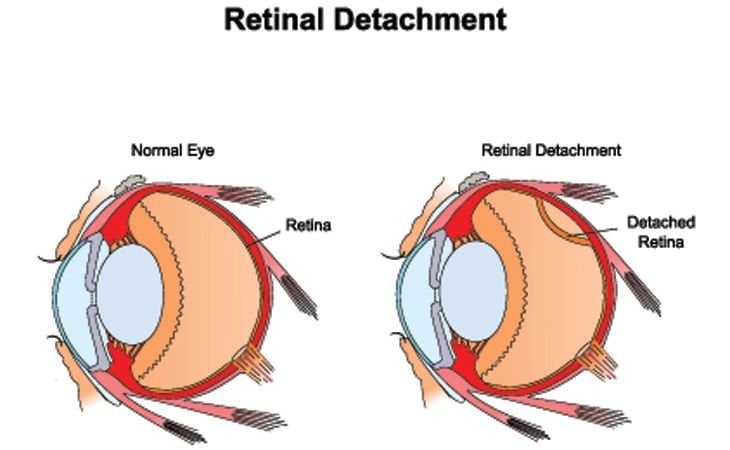
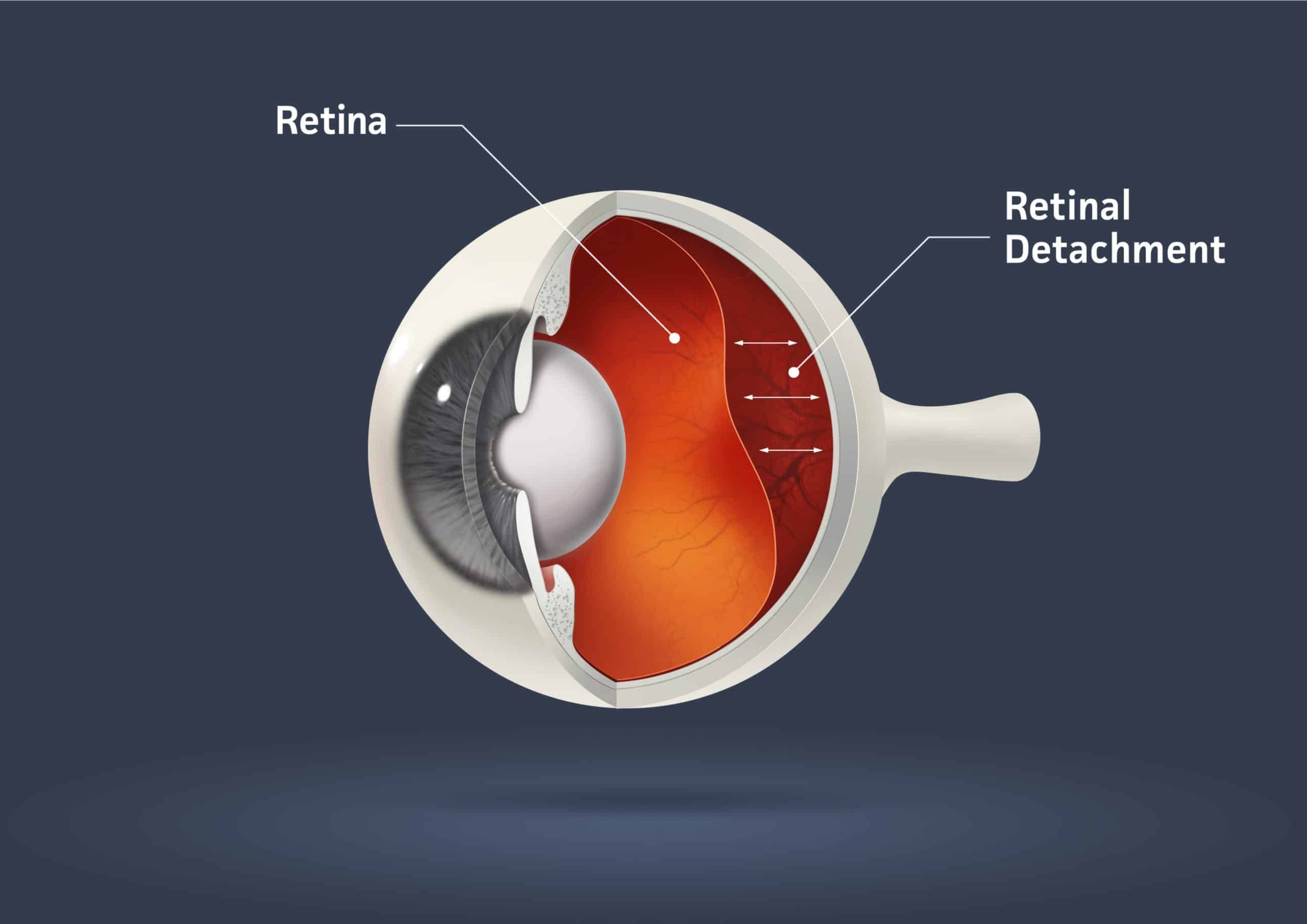 Can You Prevent and Treat Retinal Detachment?03 junho 2024
 -
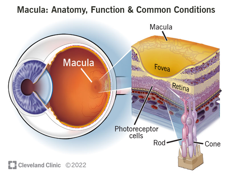
Macula: Anatomy, Function & Common Conditions03 junho 2024
-
What is a Detached Retina? Symptoms, Causes and Treatment03 junho 2024
-
 Retina Definition, Anatomy & Function - Video & Lesson03 junho 2024
 -
 Normal Eye Retina, Illustration Stock Illustration - Illustration03 junho 2024
 -
 Retina Clínica e Cirurgica - Dra. Juliana Prazeres03 junho 2024
 -
 Detached Retina, Optometrist in Chicago, Illinois03 junho 2024
 -
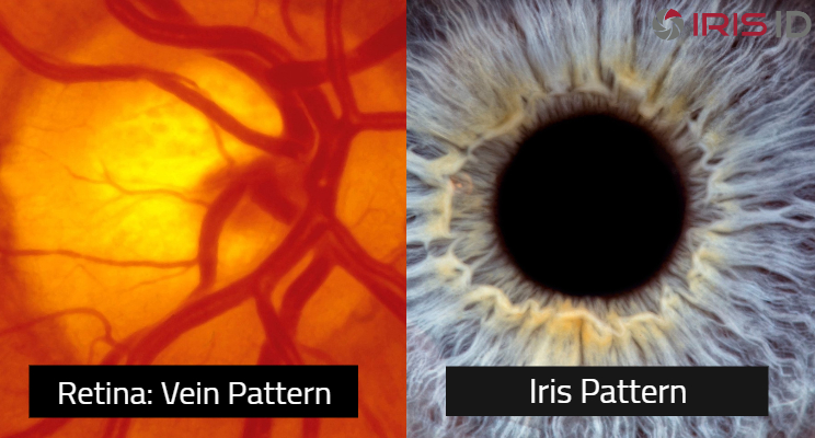 Iris recognition and retinal scans are not the same - Iris ID03 junho 2024
você pode gostar
-
 Just Friends Anna Faris GIFs03 junho 2024
 -
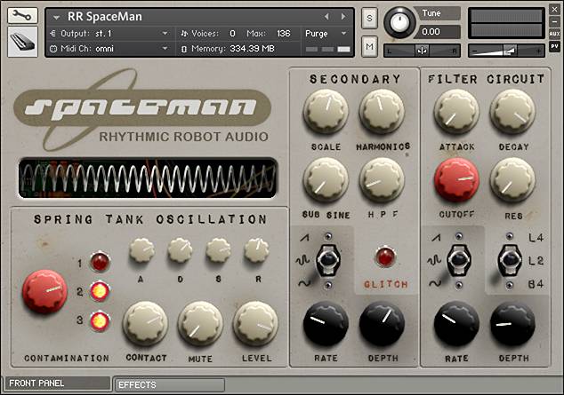 SpaceMan - vintage Kontakt synthesizer by Rhythmic Robot03 junho 2024
 -
 Gran Turismo 7 Graphics: Ray Tracing, 60 FPS, 4K resolution, gameplay, PS4, PS5, & more03 junho 2024
 -
Livros e cadernos de fichas de História do 5 ano Oiã • OLX Portugal03 junho 2024
-
 HP's Flagship OMEN 45L Gaming PC with RTX 4090 GPU Is Down to $2880 - IGN03 junho 2024
 -
 Shellshock 2: Blood Trails03 junho 2024
 -
 As casinhas de bonecas mais lindas do mercado - Mamãe Plugada03 junho 2024
 -
 Border Café (2005) - IMDb03 junho 2024
 -
 Soi kèo Arsenal Sarandi vs Independiente VĐQG Argentina 202203 junho 2024
 -
Jogo do Bicho Online added a new photo. - Jogo do Bicho Online03 junho 2024
